Exposure to one of the most toxic forms of mercury (methylmercury) may also disrupt the body’s metabolic health, according to the findings of a new international study. The research published in Chemical Research in Toxicology, found the element may have wider health effects than previously known. Methylmercury can accumulate in water environments and enter the food chain. Human exposure is of particular concern in communities affected by contaminated rivers, industrial pollution, artisanal gold mining and environmental disasters. Mercury pollution is usually associated with damage to the brain and nervous system. This new study suggests that methylmercury may also disrupt the body’s metabolic health, affecting the liver, fat tissue biology and cardiovascular disease risk in vulnerable populations.
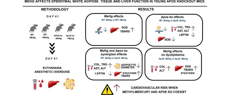
The study is a collaboration of researchers with expertise in toxicology, tissue biology, metabolism, computational modelling and advanced image analysis, from the University of Bristol (UK), the Federal University of Ceará, Brazil, and the University of California, USA. The team looked at a protein called apolipoprotein E (ApoE), which helps the body transport fats and cholesterol around the bloodstream. ApoE also plays a role in inflammation, liver health and heart disease risk. The study aimed to find out whether differences in this protein could make some people more vulnerable to the harmful effects of methylmercury. The team studied mice lacking ApoE. When methylmercury exposure was combined with ApoE deficiency, the animals showed stronger signs of metabolic disruption, including higher cholesterol and triglycerides, increased markers of liver injury, oxidative stress and changes in white fat tissue.
Dr Augusto Coppi, Senior Lecturer in Veterinary Anatomy at the University of Bristol and co-lead of the study, said: "Mercury pollution is usually viewed through the lens of neurotoxicity, but our findings suggest that its impact may reach much further. Our study indicates that methylmercury can interact with key biological systems involved in cholesterol handling, liver health and fat tissue function.
“In simple terms, the findings suggest a possible double hit: methylmercury exposure on one side and pre-existing vulnerability in lipid metabolism on the other. Together, these factors may place greater stress on organs and tissues involved in metabolic and cardiovascular health. Bristol’s expertise in 3D quantitative image analysis is enabling the team to examine tissue architecture in far greater detail — moving beyond whether organs simply look abnormal to measuring how methylmercury exposure and ApoE-related vulnerability may alter metabolically important tissues. This ongoing work will support deeper tissue-level interpretation and help clarify the study’s potential translational relevance for public health.”
Paper
- ‘In silico ApoE isoform interactions with methylmercury (MeHg) and in vivo MeHg intoxication effects on epididymal white fat tissue and liver function in young ApoE knockout mice’ by Augusto Coppi, Reinaldo B. Oria et al. in Chemical Research in Toxicology.